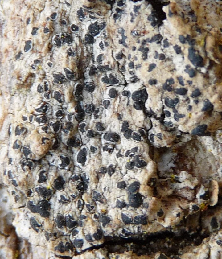
Schismatomma graphidioides
S. graphidioides is morphologically similar to several other crustose epiphytes with lirellate apothecia, e.g. Opegrapha species, but is distinct in having a raised, halo-like, rim of thalline tissue surrounding the apothecia. Being pale and usually whiter than the thallus the halo is prominent under the hand-lens.
It closely resembles Opegrapha rufescens, a species also with rimmed apothecia, from which it can be indistinguishable without microscopic examination (Smith et al., 2009; Coppins & James, 1979). Microscopically the two species differ in critical anatomical characters as detailed in the table below:
| Character | S.graphidioides | O. rufescens |
|---|---|---|
| True exciple | Absent; inapparent in sections | Narrow, thin, persistent, dark; visible in section (x100). |
| Epihymenium | Pale brown, diffuse; K-, I+ inky blue, KI+ inky blue (persistent and penetrating down 10µm or more into the hymenium) | Brown, K-, I- or slightly I+ grey or weakly bluish in places, not penetrating the hymenium. |
| Hymenium | *I+ and KI+ inky blue above, or very weak I+ orange in part | I+ deep orange-red |
| Hypothecium | Dark brown-black; K+ deep green-black, I+ and KI+ inky blue | Chestnut brown, K- (duller and lighter), I- or thin and weak I+ grey-blue at base of paraphyses in subhymenium |
| Spores | Spiralled in ascus, curved-sigmoid on release, +/- narrow-acicular, 3-sept., ca. 24 - 36(-40) x 2.8 - 3.5 µm, ends narrowed | 3-septate, fusiform, broader and shorter than in S. graphidioides, 15 - 27 x 3 - 5 µm, not spiralled, though some distinctly curved, ends rounded |
| Epihymenial crystals | With abundant (?always) cuboid to tetrahedral, oxalate crystals, ca. 4 - 7µm across, becoming more distinct and not disssolving in K | Crystals apparently absent (?always) |
| Asci | Over-mature asci frequently become brown internally from old spores | Old spores can become brownish but ascal contents rarely so and only in moribund material |
| Paraphyses | Abundant, slender, to 1.5 µm | Fewer and less distinct |
Note: *Lugol's solution Under the microscope collections are best identified by directly comparing mounted sections of verified material of either species side by side on the same slide. Spore characters and stark hypothecium and K-reaction differences are usually sufficient to effect separation. S.P. Chambers
- Read more about Schismatomma graphidioides
- Log in to post comments